| Specification |
| Catalog No. | EC-Sickle Cell 1001 | Format | Cassette |
| Specimen | Whole blood/Finer blood | Test/Box | 25T
|
| Reading Time | 10-15 minutes | Shelf Life | 3 Years |
| Storage Temperature | 4-30°C | Specificity | >99.9% |
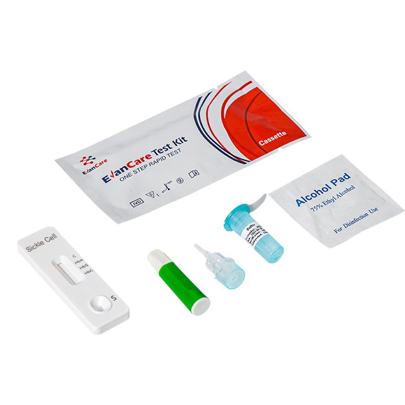
| Sensitivity | >99.9% | Composition | Individually packed test device, Package insert,Dropper,Buffer
|
| Accuracy | >99.9% | Feature 1 | High Accuracy |
| Feature 2 | High Sensitivity | Feature 3 | Clear Results |
| Feature 4 | Easy To Read | Feature 5 | Easy To Use |
| Feature 6 | OEM/ODM Available | Feature 7 | Quality Guarantee |
Test Procedure
Specimen Collection and Preparation
Use fresh Whole blood fortesting. 1.Capillary Blood:To use fresh blood from finger prick/puncture,cleanse the finger using sterile swab allow it to dry. With the help of lancet puncture the skin and collect blood in sample dispenser. 2.Venous Blood:Collect the whole blood in a blood collection tube/container having EDTA or sodium Citrate as anticoagulant. 3.Whole blood specimen should be used for testing immediately or shall be stored at 2-8℃ for up to 72 hours (3 days).Do not use blood specimen stored for more than 3 days,it can cause a non-specific reaction.
Note: Hemolytic samples should not be used! Do not freeze whole blood sample in any case.
Test Procedure
Carefully read the reagent instruction before using the test kit and strictly operate according to the instruction to ensure reliable results.
Bring test device, buffer and specimens were restored to room temperature 15-30℃(59-86℉).
Please keep the temperature at 15-30 ℃ and the humidity at 20%-80% during the whole test.
1.Remove the test from its sealed pouch, and place it on a clean, level surface. For best results, the assay should be performed within one hour.
2. Place the extraction tube on the tube rack and tear the aluminum film.
3. Pinch the bulb at the upper end of the dropper, place the lower end vertically in the blood, and then slowly loosen the bulb. The blood should be flush with the very top of the thin tube (approximately 5 uL), as shown in the figure as an example
4.Take 5μl of whole blood sample into the extraction tube and mix sample by Shaking up 10 times.
5. Dispense 2 to 3 drops(approximately 60~80μl) of mixed sample buffer to sample well of the test cassette.
6. Start the timer. Wait for the colored line(s) to appear. The test result should be read at 10 minutes. Do not interpret the result after 15 minutes.
Interpretation of test results
HbAA: Two distinct red lines appear, one in the control region (C)and another in the HA region, with no band at the HS and HC lines. This result indicates the presence of HBAA, which is considered normal hemoglobin.
HbAS: Three red lines appear, one in the control region (C),another in the HA region, and a third in the HS region, with no band at the HC line. This result indicates the presence of HBAS, which is associated with the sickle cell trait.
HbSS: Two distinct red lines appear, one in the control region (C) and another in the HS region, with no band at the HA and HC lines. This result indicates the presence of HBSS, which is associated with sickle cell disease.
HbAC: Three red lines appear, one in the control region (C), another in the HA region, and a third in the HC region, with no band at the HS line. This result indicates the presence of HBAC, which is associated with the sickle cell trait.
HbSC: Three red lines appear, one in the control region (C), another in the HS region,and a third in the HC region, with no band at the HA line. This result indicates the presence of HBSC, which is associated with a different type of sickle cell trait.
Invalid: If the control band fails to appear within the result window, the result is considered invalid. This could be due to various reasons, such as improper test procedure or deterioration of the test kit. In such cases, it is recommended to retest the specimen.
Quality Control
A red line appearing in the control region (C) is the internal procedural control. It confirms sufficient specimen volume.