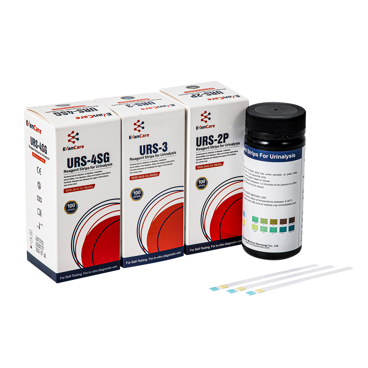
1. Remove strip from the pouch for immediate use.
2. Completely immerse reagent areas of the strip in fresh urine. Remove the strip immediately to avoid dissolving out the reagent areas.
3. While removing, touch the side of the strip against the rim of the urine container to remove excess urine. Blot the lengthwise edge of the strip on an absorbent paper towel to further remove excess urine and avoid running over (contamination from adjacent reagent pads.)
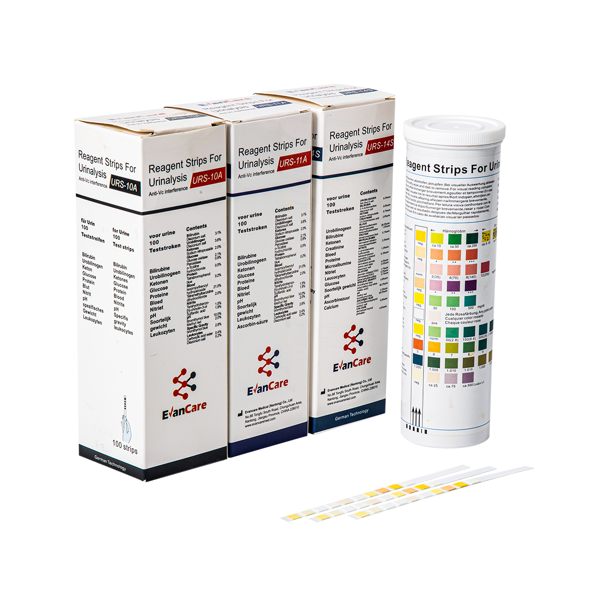
4. Compare each reagent area to its corresponding color blocks on the color chart and read at the times specified. Proper read time is critical for optimal results.
5. Obtain results by direct color chart comparison.
SUMMARY
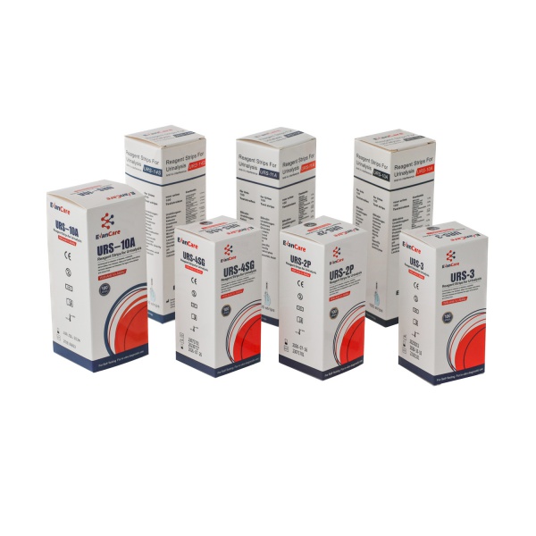
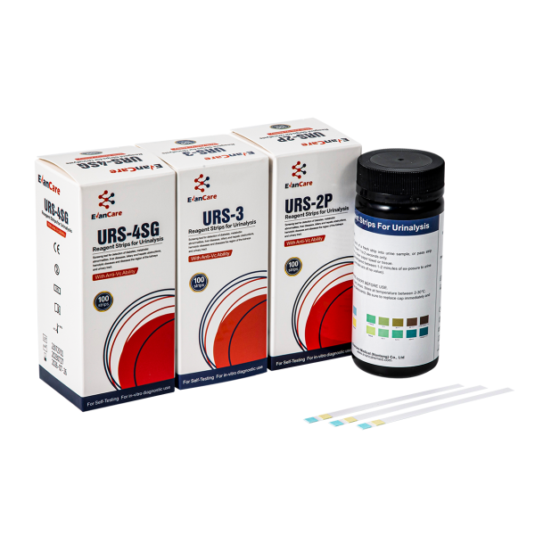
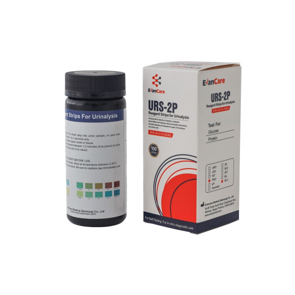
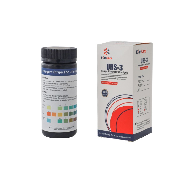
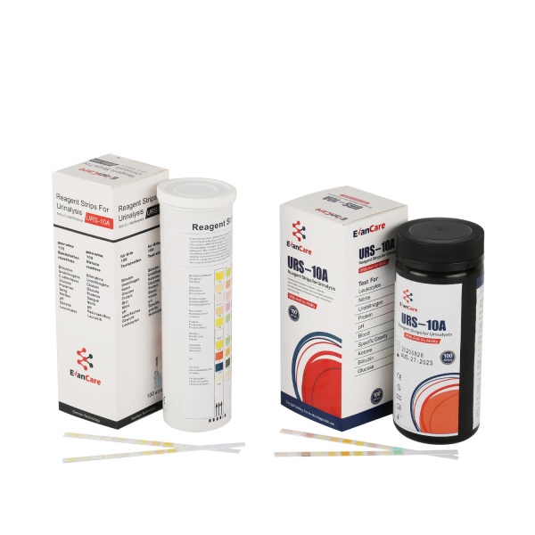
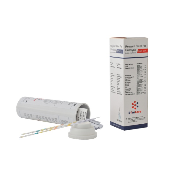
Reagent strips for urinalysis (URS) are firm plastic strips to which severaldifferent reagent areas are affixed. Depending on the product being used,Reagent strips for Urinalysis provide tests for Glucose, Bilirubin, Ketone(Acetoacetic acid), Specific Gravity, Blood, pH, Protein, Urobilinogen, Nitrite,Leukocytes, and Ascorbic Acid in Urine.
TEST PRINCIPLE
Glucose: This test is based on a double sequential enzyme reaction. Oneenzyme, glucose oxidase, catalyzes the formation of gluconic acid and hydrogenperoxide from the oxidation of glucose. A second enzyme, peroxidase, catalyzesthe reaction of hydrogen peroxide with potassium iodide chromogen to oxidizethe chromogen to colors ranging from blue to brown and dark brown. Bilirubin: This test is based on the coupling of bilirubin with a diazotizeddichloroaniline in a strongly acid medium. The colors range from turtish yellow tolight tan.
Ketone: This test is based on the reaction of acetoacetic acid with sodiumnitroprusside in a strongly basic medium. The colors range from beige or buff�pink color for a “Negative” reading to pink and pink-purple for a “Positive” reading.
Specific Gravity: This test is based on the apparent pKa change of certainpretreated polyelectrolytes in relation to the ionic concentration. In the presenceof an indicator, the colors range from dark blue or blue-green in urine of low ionicconcentration to green and yellow-green in urine of higher ionic concentration.
Blood: This test is based on the pseudoperoxidase action of hemoglobin anderythrocytes which catalyzes the reaction of 3,3’, 5, 5’-tetramethyl-benzidine andbuffered organic peroxide. The resulting colors range from light yellow to yellow�green and dark blue. Very high blood concentration may cause the colordevelopment to continue to dark blue.
pH: This test is based on the well known double pH indicator method, wherebromothymol blue and methyl red give distinguishable colors over the pH rangeof 5.0-8.5 The colors range from red-orange to yellow and yellow-green to blue-green.
Protein: This test is based on the protein error-of-indicator principle. At aconstant pH, the development of any green color is due to the presence ofprotein. Colors range from yellow for a “Negative” reaction to yellow-green andgreen to blue-green for a “Positive” reaction.
Urobilinogen: This test is based on a modified Ehrlich reaction in which p�diethylaminobenzaldehyde reacts with urobilinogen in a strongly acid medium.Colors range from light pink to bright magenta.
Nitrite: This test depends on the conversion of nitrate to nitrite by the action ofGram-negative bacteria in the urine. The nitrite reacts with p-arsanilic acid tofrom a diazonium compound in an acid medium. The diazonium compound inturn couples with 1,2,3,4-tetrahydrobenzo(h) quinolin to produce a pink color.
Leukocytes: This test is based on the action of esterase present in leukocytes,which catalyzes the hydrolysis of an indoxyl ester derivative. The indoxyl esterliberated reacts with a diazonium salt to produce a beige-pink to purple color.
Ascorbic Acid: The composition comprises of a complex chelating agent with apolyvalent metal ion in its higher state and an indicator dye that can reacts withthe metal ion in its lower state to produce a color change from blue-green to yellow.